
In emergency departments and ICUs, the multiparameter vital signs monitor is one of the most indispensable tools in medical practice. It provides continuous, real-time measurements of essential physiological variables, allowing early identification of hemodynamic and respiratory instabilities. However, for a medical student or resident, looking at a screen full of waveforms, numbers, and alarms can feel overwhelming at first.
In this Educar Med guide, we review the main components of a vital signs monitor and how to correlate each data point with your patient’s clinical examination.
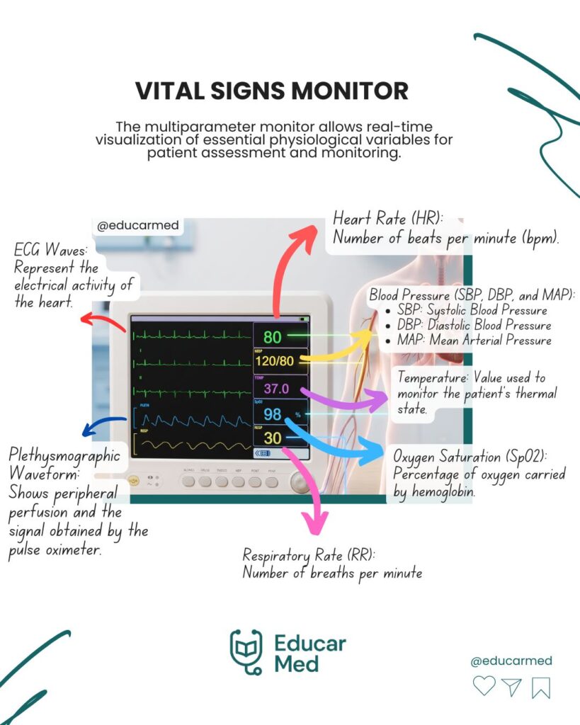
1. ECG Waveforms (Cardiac Electrical Activity)
Displays the continuous electrocardiogram tracing (usually Lead II or V5 for arrhythmia and ischemia monitoring).
- What to evaluate: Heart rhythm (sinus vs. arrhythmias), QRS complex regularity, and the presence of ST-segment elevation or depression.
2. Heart Rate (HR)
Displayed numerically (in beats per minute – bpm), derived from either the ECG tracing or the pulse oximeter.
- Adult reference range: 60 to 100 bpm.
- Red flags: Tachycardia (>100 bpm) or Bradycardia (<60 bpm).
3. Plethysmographic Waveform and Oxygen Saturation (SpO2)
The plethysmographic waveform reflects peripheral perfusion captured by the pulse oximeter. The height and shape of the waveform help assess signal reliability.
- Normal SpO2: ≥ 95% on room air.
- Clinical tip: If the plethysmographic wave is flat or irregular, the SpO2 reading may be inaccurate (common in hypothermia, vasoconstriction, or shock).
4. Blood Pressure (SBP, DBP, and MAP)
Measurement can be non-invasive (NIBP) or invasive (IBP via an arterial line, for example). The monitor displays Systolic Blood Pressure (SBP), Diastolic Blood Pressure (DBP), and Mean Arterial Pressure (MAP).
MAP Formula:
MAP = DBP + SBP – DBP / 3
Clinical goal: MAP is the primary indicator of organ tissue perfusion and should generally be maintained at ≥ 65 mmHg in most critically ill patients.
5. Respiratory Rate (RR) and Temperature
- RR: Measured in breaths per minute (brpm), typically obtained via thoracic impedance from the ECG leads.
- Temperature: Monitored via a cutaneous, esophageal, or rectal sensor, essential for screening infections or accidental hypothermia.
🚨 CLINICAL REMINDER:
Interpreting waveforms and numerical readings on a monitor should never replace a physical examination and direct clinical assessment of the patient.