
Biliary colic is one of the most common causes of acute abdominal pain in emergency rooms and outpatient clinics, especially in women of childbearing age. However, it is common to see students and newly graduated physicians underestimate the complaint, confusing the presentation with transient “indigestion,” dyspepsia, or gastritis. The problem with not investigating thoroughly is that the condition can evolve silently into severe inflammation, infections, and surgical emergencies.
In this article by Educar Med, we will integrate clinical anatomy, hepatobiliary pathology, and diagnostic management to help you refine your clinical reasoning and never miss a biliary complication.
Biliary function and stone formation
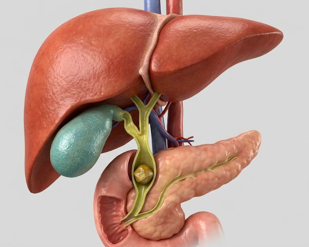
The gallbladder is a small organ located in the gallbladder fossa [lecho hepático] whose main function is to store and concentrate bile, the substance produced by the liver essential for emulsification and digestion of lipids. During gastric emptying following meals rich in fat, the release of cholecystokinin (CCK) stimulates gallbladder contraction, releasing bile into the duodenum.
Clinical reasoning starts being required when we deal with cholelithiasis (gallstone formation). Bile supersaturation with cholesterol or calcium bilirubinate leads to crystal precipitation. These stones can impact in the infundibulum of the gallbladder or the cystic duct, blocking bile outflow and causing intense pain due to distension of the gallbladder wall. Although many patients remain asymptomatic for years, transient impaction generates the classic biliary colic crisis.
Clinical presentation of biliary colic
Precise clinical recognition is the first step toward diagnostic success and rules out dangerous differential diagnoses (like acute myocardial infarction or perforated ulcer). The classic presentation includes:
- Intense visceral pain, colicky or constant, located in the right upper quadrant or epigastrium;
- Sensation of fullness or subcostal pressure;
- Clear triggering of the condition following intake of cholecystokinetic (fatty) meals;
- Associated vagal symptoms, like nausea and profuse vomiting;
- Pain with characteristic radiation to the right scapular region or ipsilateral shoulder (sign of diaphragmatic irritation).
Complications and obstructive jaundice pathophysiology
The major turning point in the physical examination is identifying when the condition has evolved from simple colic to a complex inflammatory or obstructive picture. Absolute red flags include: fever, chills, pain that does not resolve with standard analgesia, a positive Murphy’s sign, choluria (dark urine), and acholic stool (pale stool).
One of the most important clinical signs of complication is jaundice (yellowing of skin and sclera). Pathophysiologically, jaundice is a sign of hyperbilirubinemia. When a stone migrates and obstructs flow in the common bile duct (choledocholithiasis), it generates what we define as obstructive jaundice (post-hepatic).
In this scenario, bilirubin is normally conjugated by the hepatocyte but cannot reach the intestine for excretion. Consequently, it flows back into the bloodstream, elevating predominantly direct bilirubin. Persistent obstructions of this drainage pathway are the entryway to emergencies such as acute cholecystitis, severe bile duct infections (acute cholangitis), and dreaded biliary pancreatitis.
Integrated diagnosis: imaging and laboratory testing
To confirm the diagnosis and define management, the clinical aspect must reign supreme, supported by well-indicated complementary tests. The initial gold standard for investigation is complete abdominal ultrasound, which has high sensitivity for identifying stones, evaluating gallbladder wall thickening (suggesting acute inflammation), and detecting dilation of the extrahepatic bile ducts.
In the laboratory, assessing liver function is imperative to map the extent of the obstructive damage:
- Cholestasis Markers (Biliary Excretion): Flow obstruction causes enzyme induction and a marked elevation of enzymes present in the canalicular membrane: Alkaline Phosphatase (ALP) and Gamma-Glutamyl Transferase (GGT). The combined elevation confirms that the source of the problem is, in fact, the biliary tree.
- Bilirubins: Total Bilirubin elevation at the expense of Direct Bilirubin confirms the acute obstructive pattern.
- Hepatocellular Integrity: Measuring transaminases, ALT (SGPT) and AST (SGOT), helps verify if biliary reflux and retrograde pressure are already causing direct suffering to the hepatocytes.
Management and discharge directions
Clinical management depends on risk stratification and the presence of complications. Simple painful crises usually respond well to antispasmodics and NSAIDs in the ER. However, the definitive treatment for symptomatic biliary lithiasis is surgery (cholecystectomy), with laparoscopy being the current gold standard due to its lower morbidity and rapid recovery.
For the general practitioner, the discharge timing of a patient with uncomplicated biliary colic is crucial. The prescription must be accompanied by clear lifestyle change guidance to avoid returning to the surgical center on an emergency basis. Recommend:
- A diet restricted in lipids, avoiding fried foods and sausages;
- Gradual weight control strategies (very abrupt weight loss also predisposes to new stones);
- Frequent small meals to avoid biliary stasis due to long fasting periods;
- Immediate outpatient return to the General Surgery team for elective surgical scheduling.
Remember: in medicine, repeated episodes of abdominal pain should never be underestimated. Your mastery of biliary pathophysiology is what guarantees your patient’s safety.
About Educar Med
Educar Med isn’t just about passing tests; it’s about rejecting clinical mediocrity. We are a community dedicated to training the new generation of physicians who think, examine with precision, and transform lives.
Keep raising your clinical standards. Follow Educar Med on social media: